

PROTECT YOURSELF with Orgo-Life® QUANTUM TECHNOLOGY
Orgo-Life the new way to the future Advertising by Adpathway A new antibody-based drug system uses click chemistry to combine existing cancer medicines after injection. The approach may offer a faster, more flexible way to attack treatment-resistant tumors. Credit: Shutterstock
A shape-changing drug attacks tumors in several ways and improves outcomes in mice.
A tumor is rarely made up of identical cells. As cancer progresses or adapts to treatment, different cells may display different molecular targets, allowing some to escape drugs designed to recognize only one. That diversity poses a challenge for antibody-drug conjugates, precision medicines that carry a cancer-killing compound directly to cells bearing a specific receptor.
Researchers at Washington University School of Medicine in St. Louis have now modified existing antibody-drug conjugates so they can assemble inside the body and strike more than one target. In mouse models, the approach increased drug delivery to tumors and substantially improved treatment outcomes.
The findings were published in Nature.
“We’ve shown that when two cancer-targeting antibodies bind together inside the body, they accumulate at the tumor more effectively and improve treatment response,” said Patrícia M. Ribeiro Pereira, PhD, an assistant professor of radiology at WashU Medicine Mallinckrodt Institute of Radiology and a research member of Siteman Cancer Center, based at Barnes-Jewish Hospital and WashU Medicine.
“There is a lot of excitement here because we have shown that it isn’t necessary to create a whole new drug platform for each therapeutic target,” added Ribeiro Pereira. “We can repurpose antibodies that already exist to improve treatments.”
One target limits complex tumor treatment
Antibody-drug conjugates have become an increasingly important part of cancer treatment. Since 2011, 15 have been approved for cancers including leukemia and tumors of the lung, cervix and breast, often for patients whose disease no longer responds to standard chemotherapy.
Each medication contains three connected parts. A cytotoxic drug kills the cancer cell. An antibody recognizes and attaches to a receptor associated with the tumor, helping spare healthy tissue. A molecular linker joins the antibody to its toxic cargo.
This precision is also a limitation. Because the drug is paired with a single antibody, it attacks only cells carrying the matching receptor. That can work well against relatively uniform tumors, but it becomes less effective when a tumor contains several cell populations with different targets.
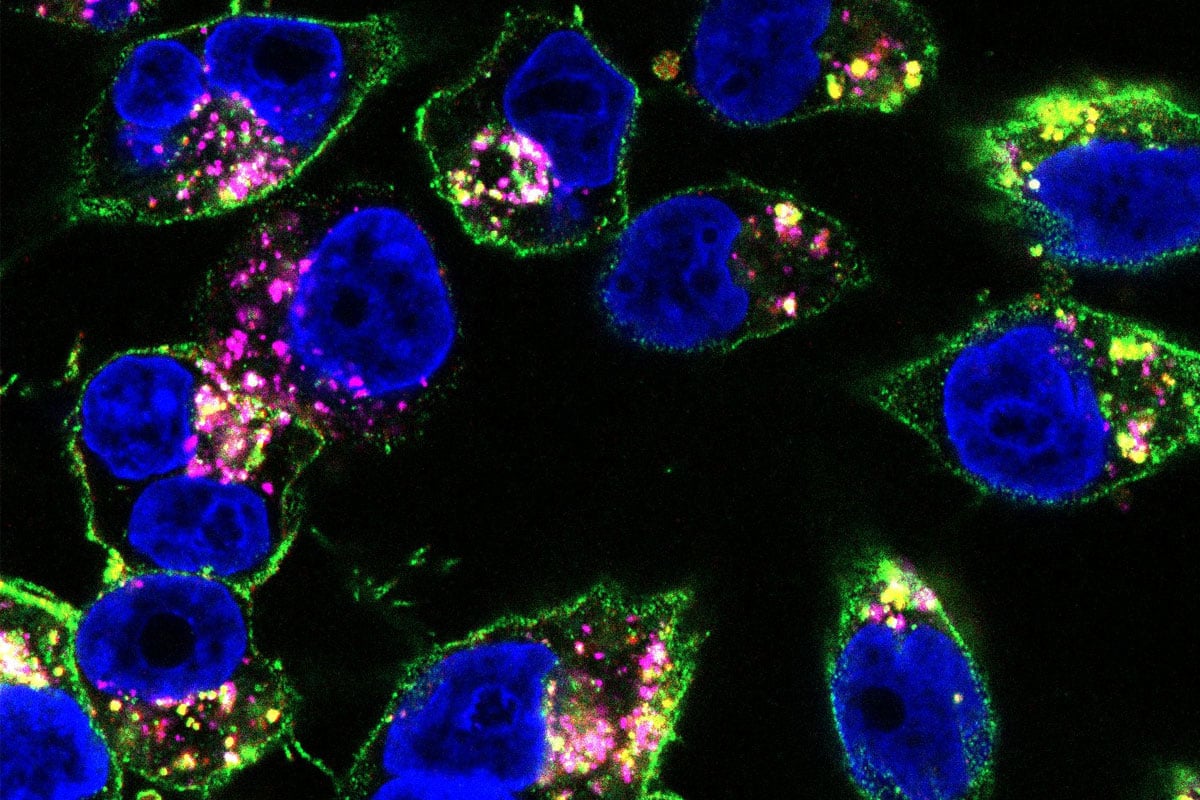 WashU Medicine researchers show in mice that their technique to designing medications, in which modular components of anti-cancer drugs are administered separately and assembled in the body using what’s known as click chemistry, is more effective than standard treatments at targeting and shrinking tumors. Shown is a drug they designed, labeled in pink and green, bound to pancreatic cancer cells from mice. Credit: Shayla Shmuel
Click chemistry adds another target
Ribeiro Pereira and her colleagues addressed that problem with click chemistry, a method that allows specially designed molecular components to connect quickly and selectively. They used it to build a modular treatment in which one antibody-drug conjugate could join with a second antibody after both had entered the body.
Adding another antibody expanded the number of tumor receptors the treatment could recognize. Rather than redesigning an entirely new drug for every combination of targets, the researchers could adapt antibodies already used in approved cancer therapies.
The study focused on two receptors involved in tumor growth, EGFR and HER2. One antibody recognized EGFR, while another targeted HER2. The researchers also tested a version that paired two different HER2 antibodies, allowing them to attach to separate regions of the same receptor and work together.
Self-assembly boosts tumor delivery
The researchers evaluated the strategy in mice with pancreatic, gastric or breast tumors containing a mixture of cells expressing EGFR and cells expressing HER2.
First, they administered either an EGFR-targeting antibody or a HER2 antibody that bound to one section of the receptor. Each had been equipped with one half of a complementary click chemistry pair.
About 24 hours later, the mice received another HER2 antibody that carried both a cancer-killing drug and the matching click component. This second antibody attached to a different region of HER2. Once the antibodies reached the body, their complementary connectors brought them together selectively.
The resulting structure could attack HER2 at two sites or recognize both HER2 and EGFR at the same time. In both cases, the assembled treatment proved substantially more effective than the FDA-approved antibody-drug conjugates from which it was created.
Radioactive labels developed by Ribeiro Pereira’s WashU Medicine colleagues allowed the researchers to track how much of the treatment reached the tumors. The modified conjugates accumulated inside cancer cells at considerably higher levels than the original drugs.
One possible explanation is that click chemistry caused antibodies to cluster on the surface of tumor cells. That clustering may have encouraged the cells to pull more of the drug inside, increasing delivery of the toxic payload.
The survival results were especially pronounced in the pancreatic cancer model. As many as 90% of treated mice remained alive 120 days after receiving the modified therapy. By comparison, mice given standard antibody-drug conjugates survived for less than 80 days on average.
The researchers also adjusted the system to reduce unwanted drug accumulation in the liver, an important consideration for limiting effects outside the tumor.
The platform could reach harder diseases
Although the experiments involved pancreatic, gastric and breast cancer models, Ribeiro Pereira believes the modular system could potentially be adapted to many other tumors and perhaps diseases that remain difficult to treat with conventional medicines.
The molecular connectors require only one to three days to manufacture. Their adaptability could allow researchers to combine different approved antibodies according to the targets present in an individual patient’s cancer, creating more flexible precision treatments without rebuilding the platform each time.
“We’re trying to optimize this tool to help antibodies reach tumors that are normally very difficult to treat, such as brain tumors,” Ribeiro Pereira said. “It’s exciting, because the drug development process doesn’t need to start from the beginning — we can use drugs that are already FDA-approved, which could help bring improved treatments to the clinic more quickly. At the same time, the approach is flexible enough to be adapted to new cancer targets as we learn more about what drives treatment resistance.”
Reference: “Modular in vivo antibody–ADC click to reverse drug resistance in tumours” by Cristina Simó, Alexander C. Vanover, Ricardo D’Oliveira Albanus, Sandeep Surendra Panikar, Shayla Shmuel, Alex Benton, Jader Giraldo-Guzman, José M. Luna, Yifei Xu, Na-Keysha Berry, Nai Keltee, Jingxia Liu, Farrokh Dehdashti and Patrícia M. R. Pereira, 15 July 2026, Nature.
DOI: 10.1038/s41586-026-10789-w
Research reported in this publication was supported by the National Cancer Institute of the National Institutes of Health (R37CA276498 and R21CA291680), internal funds provided by the Mallinckrodt Institute of Radiology, and the American Cancer Society (IRG-21–133–64–03) and the Breast Cancer Alliance. Further support came from the Alvin J. Siteman Cancer Center through The Foundation for Barnes-Jewish Hospital and the National Cancer Institute (P30 CA091842). Other support came from the W. M. Keck Foundation and the American Cancer Society Award (PF-25-1515996-01-PFCDET), National Institutes of Health (K99AG086583), a Gates Sr. Alzheimer’s Disease Research Fellowship from the Alzheimer’s Disease Data Initiative, the National Cancer Institute of the National Institute of Health under Award Number K22CA282357. The Preclinical Imaging Facility was supported by NIH/NCI Siteman Cancer Center (SCC) Support Grant P30CA091842, NIH instrumentation grants S10OD018515 and S10OD030403, and internal funds provided by the Mallinckrodt Institute of Radiology. TEM and confocal experiments were supported by the Washington University School of Medicine, The Children’s Discovery Institute of Washington University, and St. Louis Children’s Hospital (CDI-CORE-2015-505 and CDI-CORE-2019-813) and the Foundation for Barnes-Jewish Hospital (3770 and 4642).
Never miss a breakthrough: Join the SciTechDaily newsletter.
Follow us on Google and Google News.





















 English (US) ·
  French (CA) ·
  French (FR) ·