

PROTECT YOURSELF with Orgo-Life® QUANTUM TECHNOLOGY
Orgo-Life the new way to the future Advertising by Adpathway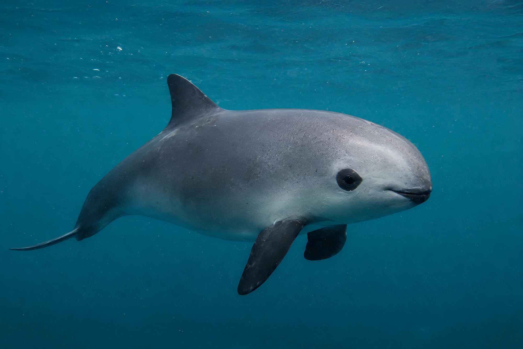 The vaquita is the world’s smallest porpoise, growing to about 5 feet long and recognizable by its dark eye rings, dark lips, and compact gray body. Found only in Mexico’s northern Gulf of California, it is critically endangered, with only a tiny number believed to remain in the wild after decades of accidental entanglement in gillnets. Credit: Shutterstock
Researchers have turned a fragile vaquita skeleton into freely accessible 3D models, preserving detailed anatomy as the porpoise nears extinction.
Only an estimated handful of vaquitas (Phocoena sinus) remain in the shallow waters of Mexico’s northern Gulf of California, the only place this small porpoise lives. With the species nearing extinction, researchers have created a way for people to examine its skeleton in detail without touching a fragile bone.
A team from Florida Atlantic University, working with the San Diego Natural History Museum, SeaWorld California, and NOAA Fisheries, digitized a complete female skeleton donated to the museum in 1966. Their study, published in Marine Mammal Science, describes one of the most comprehensive digital anatomical records of a vaquita ever assembled.
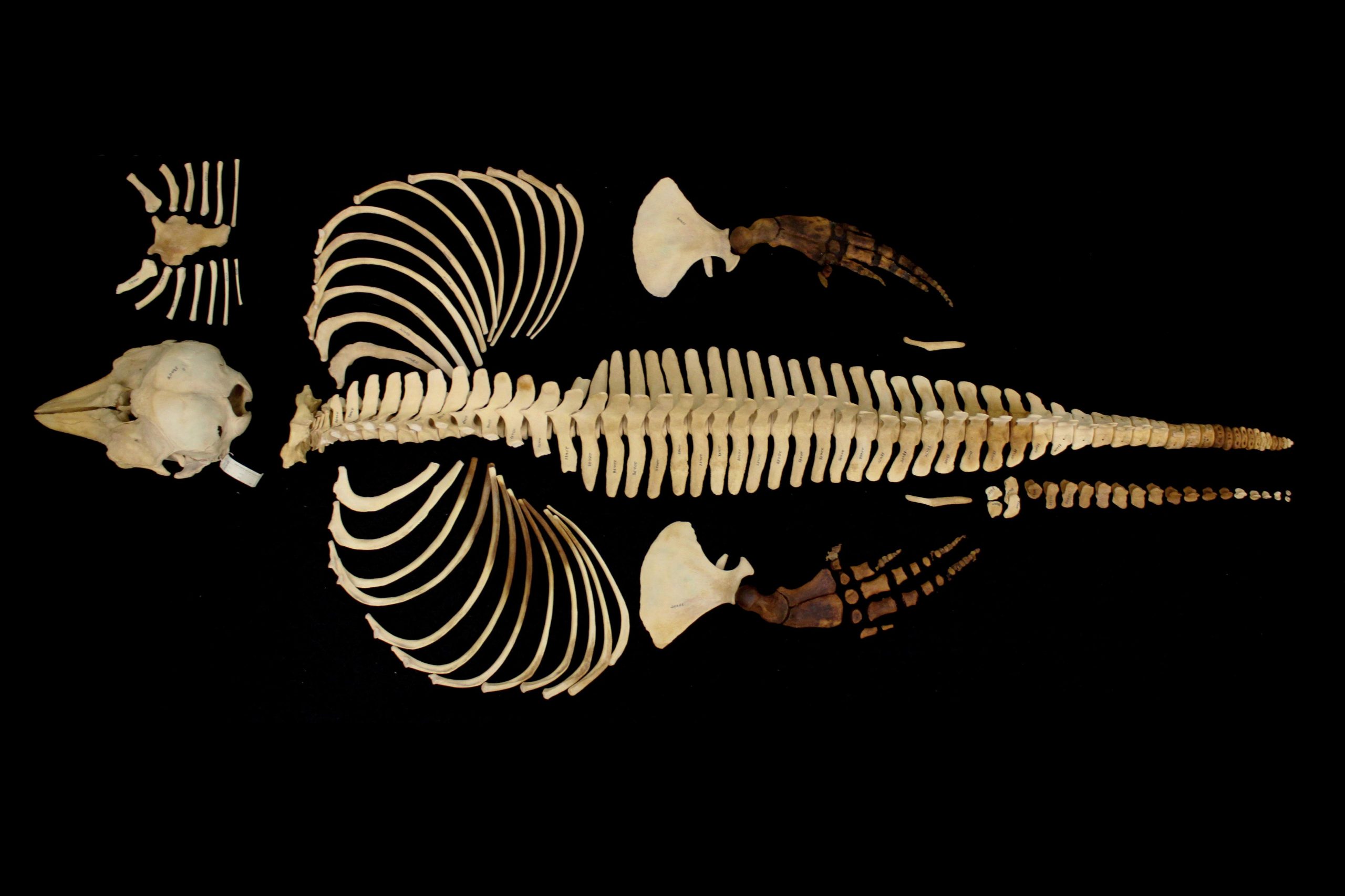 Full skeleton of a very rare vaquita specimen from the 1960s. The completed scans, which required approximately 165 hours, resulted in a total of three terabytes of data. Credit: Jamie Knaub, Florida Atlantic University
A vaquita skeleton anyone can examine
The original specimen is so rare and delicate that handling and public access are tightly limited. Its digital counterparts, now freely available through the online repository MorphoSource, can be rotated, enlarged, and examined from any angle without risking damage to the bones.
The skeleton belongs to the world’s smallest cetacean, the group that includes whales, dolphins, and porpoises. Vaquitas measure about 5 feet (1.5 meters) long and have distinctive dark rings around their eyes and mouths. These elusive animals remained unknown to science until the latter half of the 20th century.
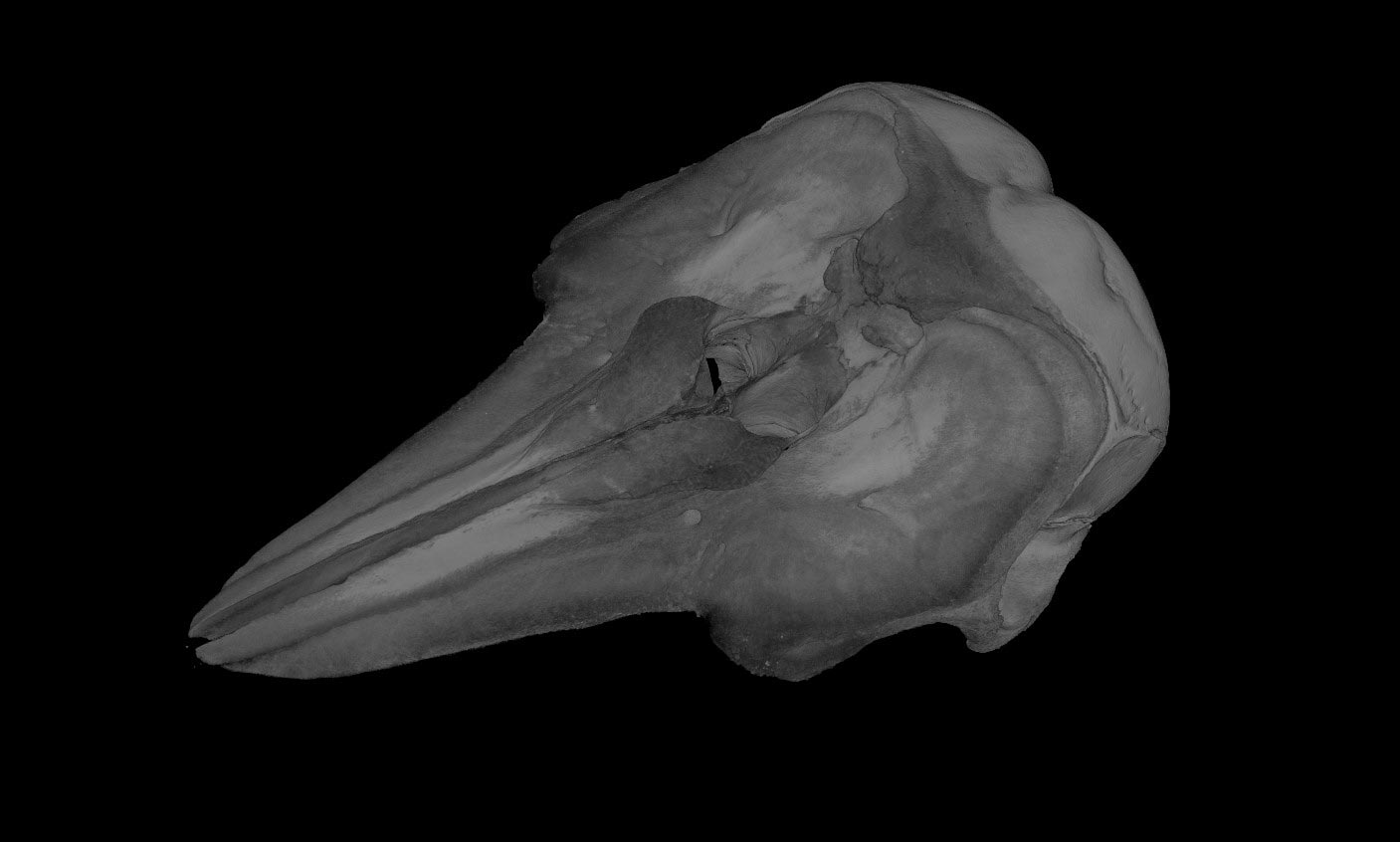 Volume rendering of the skull of a very rare vaquita specimen from the 1960s. Credit: Jamie Knaub, Florida Atlantic University
One of the study’s co-authors, Robert L. Brownell Jr., Ph.D., collected the specimen in the 1960s while he was a graduate student. He is now a biologist with the Marine Mammal and Turtle Division of NOAA Fisheries’ Southwest Fisheries Science Center. Brittany Aja Dolan, formerly with SeaWorld California, spearheaded the digitization project, working with Brownell, Philip Unitt, curator of birds and mammals at the San Diego Natural History Museum, and first author Jamie Knaub.
Gillnets leave vaquitas near extinction
In the decades since the skeleton was collected, accidental entanglement in gillnets has driven the vaquita almost to extinction. The danger comes particularly from nets set illegally for totoaba, a large fish whose swim bladder commands high prices on international black markets. Although the totoaba fishery was banned decades ago, organized wildlife trafficking networks and strong overseas demand keep illegal fishing going.
Intensive conservation efforts have left the vaquita’s future uncertain. Its survival depends on urgent, coordinated international action to eliminate gillnet fishing in its habitat. The porpoise has become a global symbol of the biodiversity crisis in the oceans, and the researchers hope the digital archive will help more people learn about it.
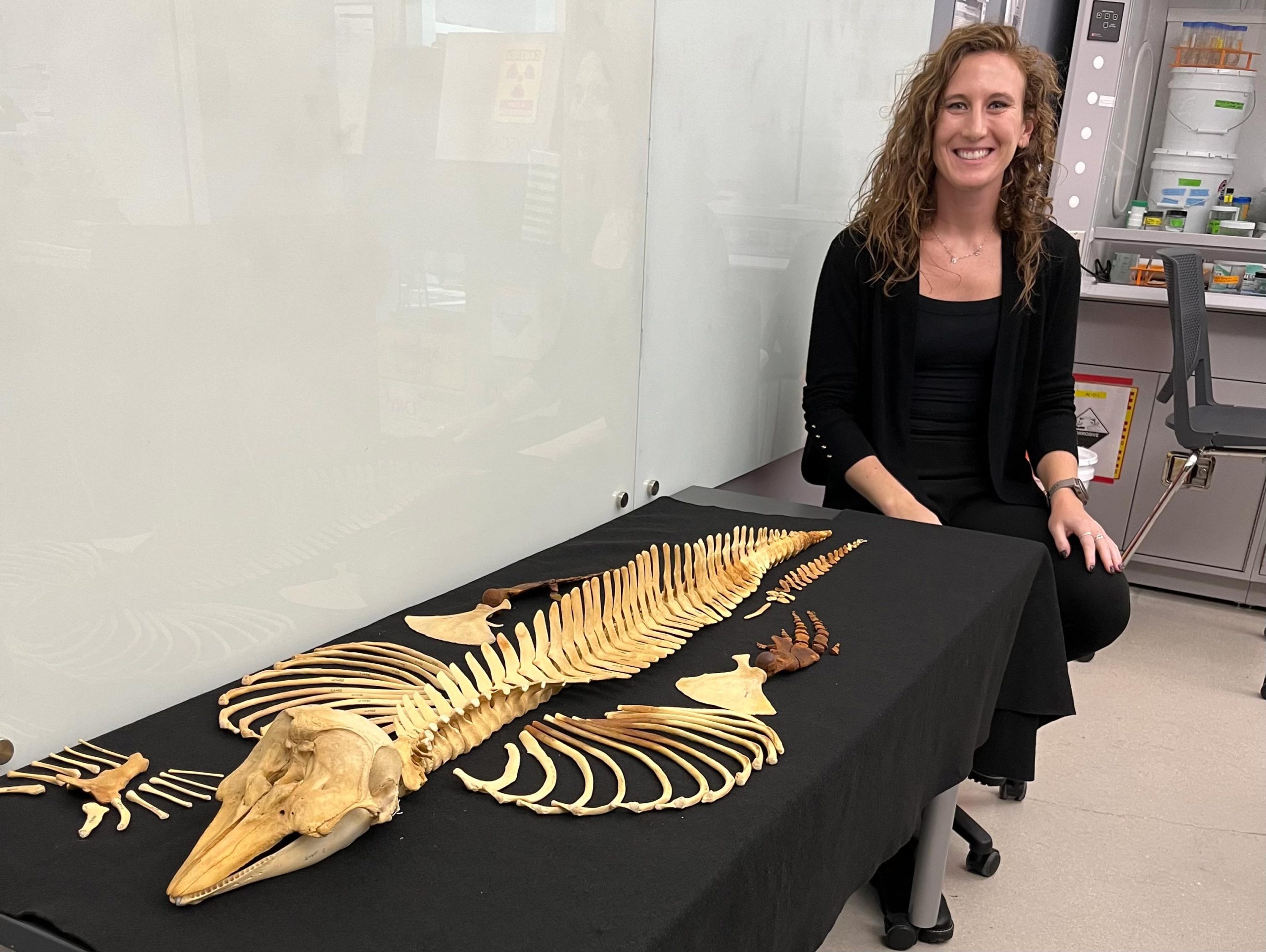 Jamie Knaub pictured with the full skeleton of a very rare vaquita specimen from the 1960s. Credit:
Tricia Meredith, Ph.D., Florida Atlantic University
“By combining advanced imaging technologies with open-access data sharing, the effort not only safeguards a valuable record of one of the planet’s most endangered marine mammals, but also makes that information accessible to anyone,” said Knaub, an imaging lab assistant in the Berlin Family Bioimaging Lab at FAU Laboratory Schools Marcus Research and Innovation Center and a Ph.D. candidate in the FAU Department of Biology within the Charles E. Schmidt College of Science. “The project will enable the production of scientifically accurate replicas for museums, classrooms, and educational programs, helping to raise awareness and support conservation efforts for a species now teetering on the edge of extinction.”
A microscopic view inside vaquita bones
Building the archive required imaging the skeleton at several scales. The researchers began with medical computed tomography, or CT, which uses X-rays to produce cross-sectional images and is commonly used in hospitals. They then photographed individual bones and skeletal elements before scanning them with high-resolution micro-computed tomography, or micro-CT.
Micro-CT captures much finer structures than conventional medical CT, revealing details measured in microns, smaller than the width of a human hair. Together, the scans generated thousands of cross-sectional images of the bones.
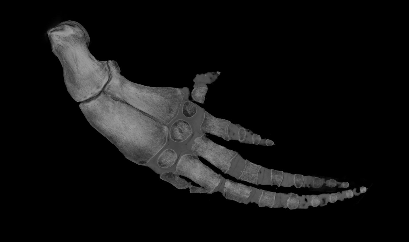 Volume rendering of the flipper of a very rare vaquita specimen from the 1960s. Credit: Jamie Knaub, Florida Atlantic University
“The success of this project was made possible by the advanced imaging capabilities available at the Berlin Family Bioimaging Lab,” said Tricia L. Meredith, Ph.D., a co-author and director of research for FAU’s on-site lab schools, A.D. Henderson University School and FAU High School, and an assistant research professor in FAU’s College of Education. “Having access to high-resolution micro-CT systems, alongside the expertise to process and reconstruct large, complex datasets, was essential to transforming raw scan data into usable 3D models. This kind of integrated technological environment is what allows specimens like the vaquita to be preserved and shared at a level of detail that simply wasn’t possible until recently.”
Using specialized 3D imaging software, the team digitally separated each bone from the scan data and reconstructed it as a detailed model. The archive retains information from the different imaging methods, allowing researchers to examine features at scales that a single scan could not capture.
“This project required an unusually intricate imaging workflow to capture the vaquita skeleton at multiple scales, from whole-bone structure down to microscopic internal detail,” said Marianne E. Porter, Ph.D., senior author and a professor in FAU’s Department of Biological Sciences. “By integrating medical CT, micro-CT, and high-resolution photography, we were able to reconstruct both the external morphology and internal architecture of each bone in a way that preserves anatomical fidelity while remaining fully interactive in digital form. The result is not just a model, but a layered dataset that reflects the true complexity of the specimen.”
Reference: “Preserving an Imperiled Porpoise Through Pixels: Digitization of a Vaquita (Phocoena sinus) Skeleton, the World’s Most Endangered Marine Mammal” by Jamie L. Knaub, Brittany A. Dolan, Philip Unitt, Robert L. Brownell, Tricia L. Meredith and Marianne E. Porter, 15 May 2026, Marine Mammal Science.
DOI: 10.1111/mms.70162
The research was supported by FAU’s School of Environmental, Coastal, and Ocean Sustainability (ECOS), the Joshua M. Berlin Research Gift, FAU Laboratory Schools, and SeaWorld California.
Never miss a breakthrough: Join the SciTechDaily newsletter.
Follow us on Google and Google News.







 English (US) ·
  French (CA) ·
  French (FR) ·